Anatomy Of Chest X Ray : Thorax Radiologic Anatomy / Both lungs should be well expanded and similar in volume.. Abcde aproach the anatomy of the heart can appear artificially larger due to this image orientation. Published 2011 by blackwell publishing ltd. A method for examining a chest. Is there any inhaled foreign body? Look for lung and pleural pathology.
Chest X Ray Of A Patient With History Of Pleural Effusion Bmj Case Reports from casereports.bmj.com Xray is a type of radiography and most widely used investigation. Conclusion of living anatomy of the chest congratulations! Posted by radiologypics ⋅ march 17, 2013 ⋅ leave a comment. • the straight back syndrome or pectus. It is almost always the first imaging study ordered to evaluate for pathologies of the thorax, although further diagnostic imaging, laboratory tests. It is used to evaluate the lungs, heart and chest what are the limitations of chest radiography? Doctors use them to diagnose problems. The interpretation of a chest film requires the understanding of basic principles.
Air spaces normally seen in.
L the portion of the left lung that corresponds anatomically to the right middle lobe is incorporated into the left upper lobe. Abcde aproach the anatomy of the heart can appear artificially larger due to this image orientation. It is used to evaluate the lungs, heart and chest what are the limitations of chest radiography? The interpretation of a chest film requires the understanding of basic principles. • the straight back syndrome or pectus. A collection of anatomy notes covering the key anatomy concepts that medical students need to learn. In this article we will focus on: Published 2011 by blackwell publishing ltd. Is there any inhaled foreign body? Therefore, knowing the basics and pathologies in the ed setting is very important. Each of these anatomical structures should be viewed using a systematic approach. It first appears too complicated to read the chest xrays because we barely know what. Many clinical conditions can be evaluated by this simple radiology test.
A method for examining a chest. Therefore, knowing the basics and pathologies in the ed setting is very important. Patterns in 21st century lung infections. It first appears too complicated to read the chest xrays because we barely know what. Both lungs should be well expanded and similar in volume.
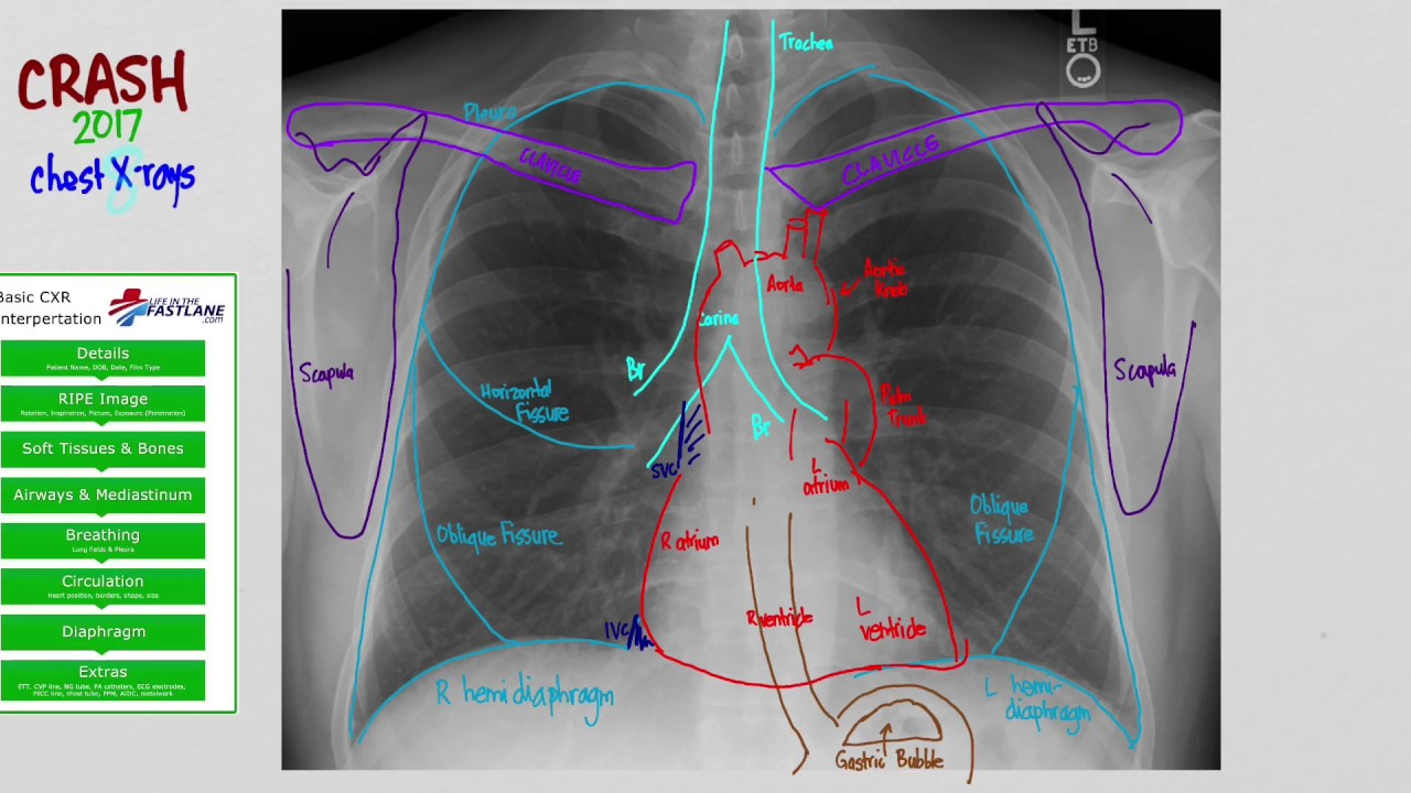
Chest X Ray Heart Anatomy My Own Rad from ronaldswanger.files.wordpress.com There are also important structures that are obscured or become visible. Because some conditions of the chest. In fact every radiologist and pulmonary physician should be an expert in chest film reading. You have completed this module. It first appears too complicated to read the chest xrays because we barely know what. Common symptoms that can be diagnosed using chest. A method for examining a chest. Each of these anatomical structures should be viewed using a systematic approach.
Patterns in 21st century lung infections.
Labeled chest radiographs teaching radiologic anatomy with a level of detail appropriate for medical students. In this article we will focus on: Living anatomy of the chest for 1st year medical students original version compiled by dr. Each of these anatomical structures should be viewed using a systematic approach. Gillian lieberman forthe harvard 62. Published 2011 by blackwell publishing ltd. The interpretation of a chest film requires the understanding of basic principles. Xray is a type of radiography and most widely used investigation. It is almost always the first imaging study ordered to evaluate for pathologies of the thorax, although further diagnostic imaging, laboratory tests. Many clinical conditions can be evaluated by this simple radiology test. A collection of anatomy notes covering the key anatomy concepts that medical students need to learn. In fact every radiologist and pulmonary physician should be an expert in chest film reading. In fact every radiologst should be an expert in chest film reading.
Therefore, knowing the basics and pathologies in the ed setting is very important. L the portion of the left lung that corresponds anatomically to the right middle lobe is incorporated into the left upper lobe. Living anatomy of the chest for 1st year medical students original version compiled by dr. Because some conditions of the chest. • the straight back syndrome or pectus.
01 Cxr Anatomy Youtube from i.ytimg.com • the straight back syndrome or pectus. Common symptoms that can be diagnosed using chest. Chest radiographs are the most common film taken in medicine. The interpretation of a chest film requires the understanding of basic principles. Labeled chest radiographs teaching radiologic anatomy with a level of detail appropriate for medical students. It first appears too complicated to read the chest xrays because we barely know what. L these two lobes are separated by a major fissure, identical to that seen on the right side, although often slightly more inferior in location. Each of these anatomical structures should be viewed using a systematic approach.
In fact every radiologist and pulmonary physician should be an expert in chest film reading.


0 Comments